Laminitis in Depth
The Anatomy of the Hoof
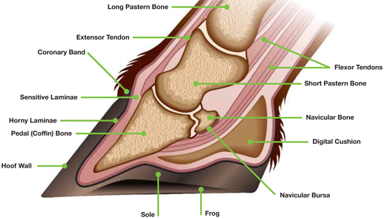
The laminae are thin tissue in the hooves of horses. The hoof of a horse is a very complicated, living structure. The outermost layer of the hoof (or the hoof wall) is the only visible portion of the horse's hoof. It is a keratinized structure very similar to our fingernail. The horse's coffin bone (also known as the distal phalnax or third phalnax) is like the bone underneath our fingernail. It fits into a horse's hoof smoothly and is attached to the inside of the hoof wall with the laminae. There are two types of laminae: the sensitive (or dermal) laminae, and the insensitive (or epidermal) laminae. The epidermal laminae make up the innermost portion of the hoof wall and cover the dermal laminae. The epidermal laminae are non-living, except for the lowest level, which generates new cells (called keratinocytes) that are dependent upon the circulation of the dermis below for nutrition. The epidermal and dermal laminae are attached by a structure called the basement membrane (or dermoepidermal junction). The epidermal laminae have an interior layer of cells called the laminar basal epithelial cells, which lie at the interface of the epidermal and dermal laminae.
The Causes of Laminitis
There are three types of laminitis and each type has different causes. The types are listed below.
Endocrinopathic Laminitis: caused by metabolic disorders like Cushing's Disease. It also happens when horses overload on carbohydrates by eating too much grain, or when obese horses eat very rich grass high in sugars in the spring or fall. It is important to do blood tests on these horses to check insulin levels.
Inflammatory or Sepsis-Related Laminitis: happens when the horse becomes systemically ill or inflamed caused by a body-wide illness or sepsis. This is the only type of laminitis that researchers have found to have a strong link to inflammation.
Weight-Bearing Laminitis: happens when a horse has an injury in one leg and is forced to bear too much weight for too long on the opposite leg.
What Laminitis Does
Laminitis causes the laminae to release their hold on the hoof wall and displace the coffin bone. The coffin bone can either rotate, sink, or tip to one side. In severe cases, the coffin bone can sink and rotate, sometimes poking through the sole of the hoof. The permanent displacement of the coffin bone is called founder.
The Beginning of Laminitis
Researchers currently think that laminitis starts because of a certain biomedical process in the hoof. The basal epithelial cells on the epidermal laminae attached to the basement membrane release when there is an injury to the hoof wall and migrate through the epidermal laminae to the hoof wall to heal the injury. This is able to happen because these epithelial cells can release enzymes called matrix metalloproteinases (MMPs) that can break down their connection with the basement membrane and the membrane itself. Normally, these enzymes are under strict control. However, when things go wrong in the horse's hoof and with the biomedical signals that keep MMPs under control, the horse could end up with a sudden overproduction/abundance of these enzymes. As a result, all of the MMPs break down the basement membrane, causing a weakening of the connection between the dermal and epidermal laminae. This weakening lets the basal epithelial cells lose their hold and laminitis sets in. Researchers currently do not know what goes wrong and why this happens. It is currently a major research problem in the horse world.
The Developmental Stage
Laminitis starts with a developmental stage that can last from 24 to 40 hours. During this stage of the disease, it is very hard to catch and treat because the horse displays no signs or symptoms. Our technology would be a preventive and diagnostic device. Owners of horses at risk for laminitis would check their horses weekly with our device, while owners of horses not at risk would only need to check every month or every two months. The L.E.A.D.S. would be able to practically enter the horse's hoof using advanced radiography and x-ray technology and spot the disease at the beginning of the developmental stage. This would allow the treatment to begin much earlier, possibly saving the horse's life.