Key |
Prototype |
|
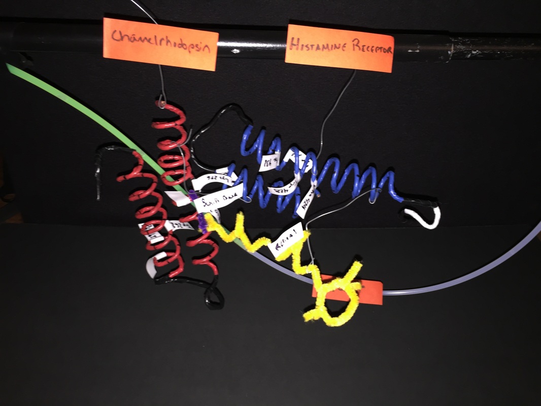
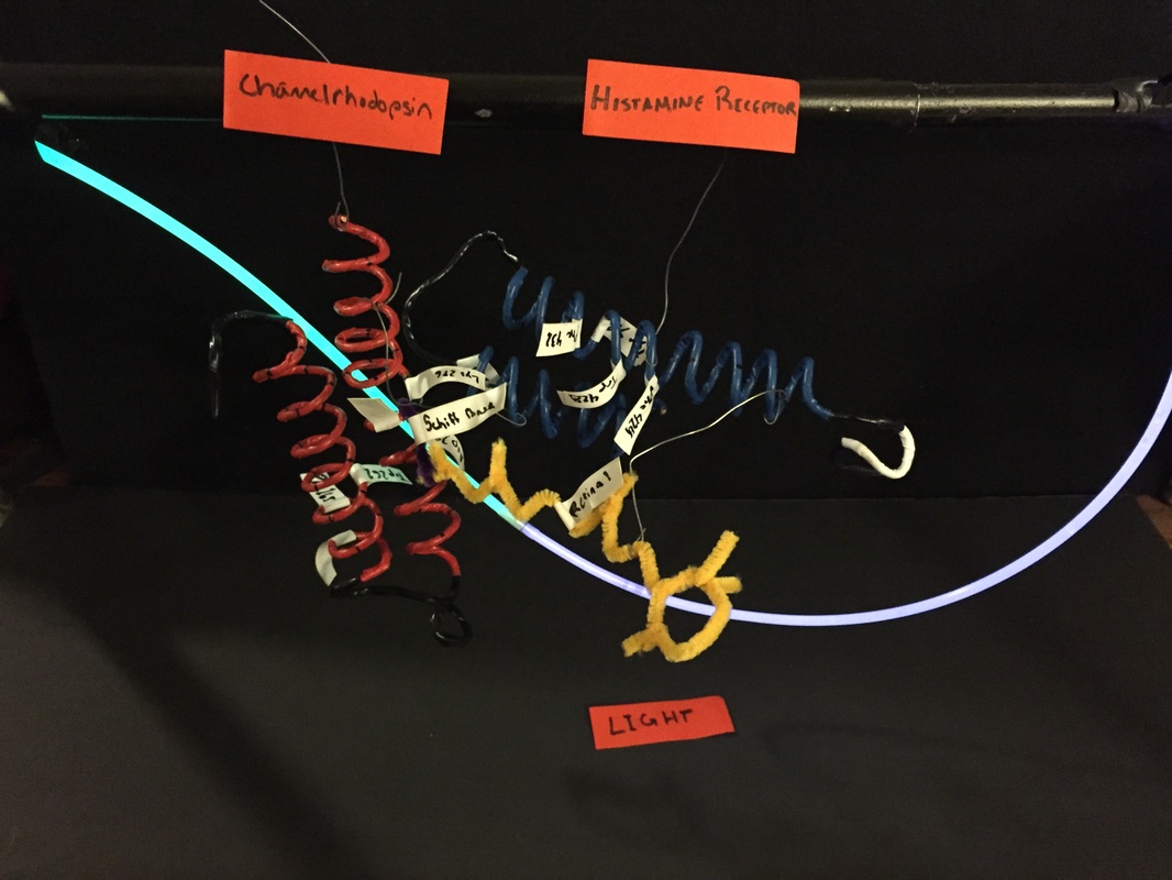
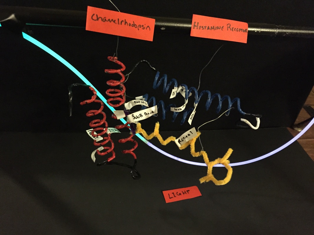
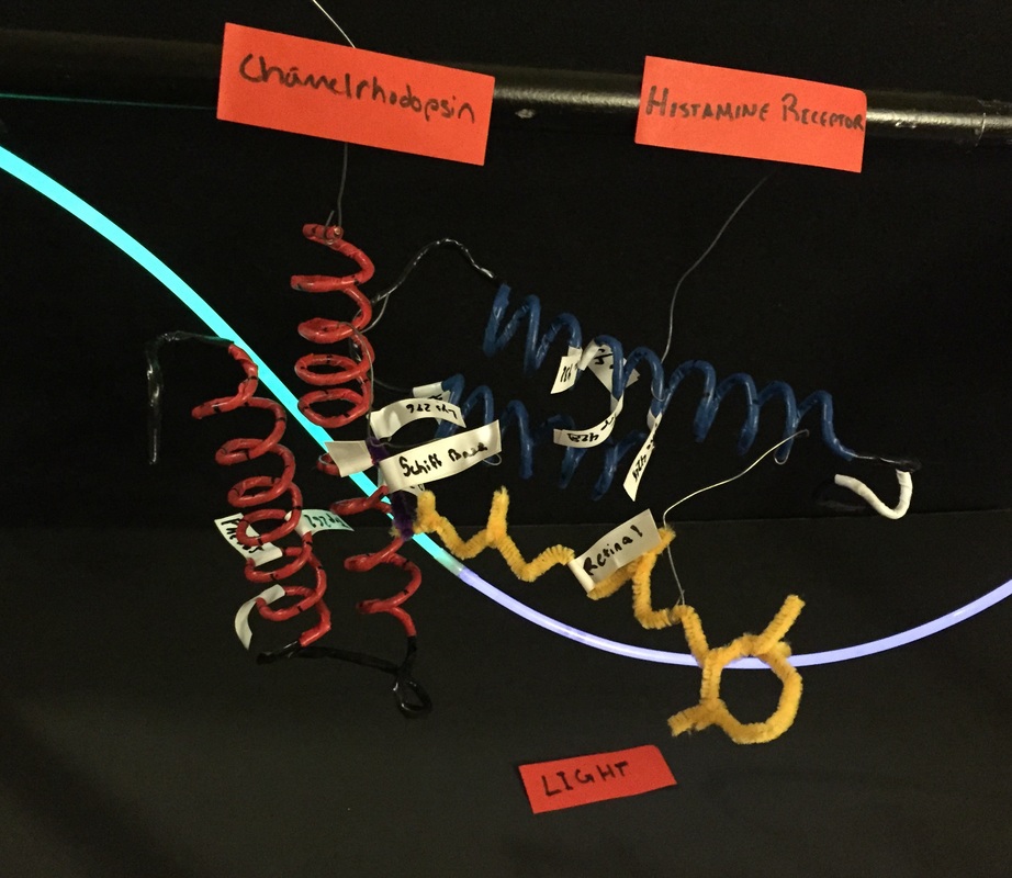
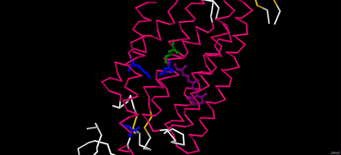
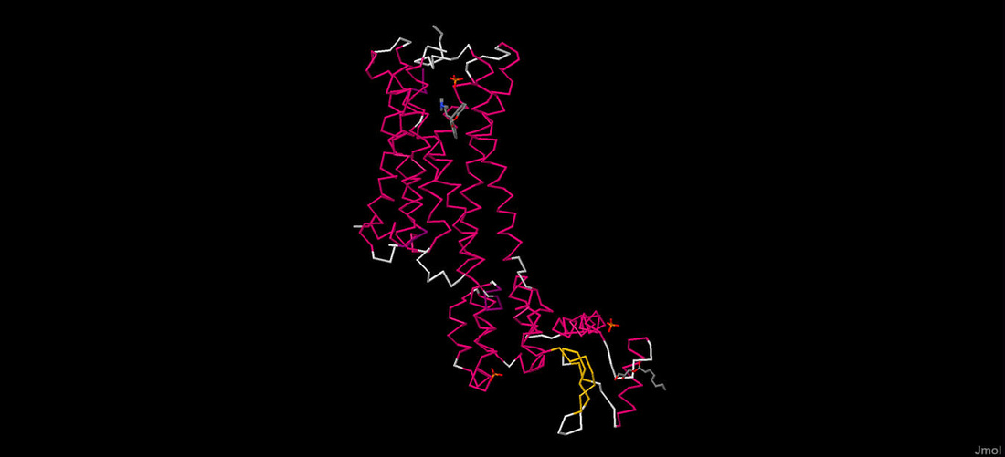
The red and blue sections of the protein model represents the alpha helix of a protein. The protein depicted with red alpha helices is channelrhodopsin, whereas the one with blue alpha helices is Histamine-1 Receptor. The black sections of the protein model are parts of the protein that don't fit into the typical scheme of the secondary structure. The white section on the Histamine 1 Receptor is a 3-10 Helix. The yellow extension of the protein is a the substrate Retinal. All tags on the protein are labels for residues at specific points. The light source in the background represents a fiberoptic within the brain shining light onto the proteins.
|
The prototype is a zoomed in protein model of Channelrhodopsin interacting with a model of Histamine 1 Receptor and a model of Retinal. In the prototype, the light source is depicted as affecting the schiff base (labelled on the model) and causing an ion to move and connect with the H1-Receptor.
|
Channelrhodopsin
|
Histamine 1 Receptor
|
The Jmol Computer Simulation
The prototype model is based off of a computer software, Jmol, which processes data detailing the structures of proteins. In the prototype model, segments of each of the computer simulations depicted above were molded to depict the interaction between the two proteins and the light source. In the side tabs on the drop down menu for the tab titled "prototype" there are links to an interactive online setting for a 3D simulation of each of these proteins.